|
Featured Instruments in the NEST Laboratory To schedule time on any of these instruments, please visit the Calendar/Scheduler section of this site. User Guides for the instruments can be found on our User Guide Index Page.
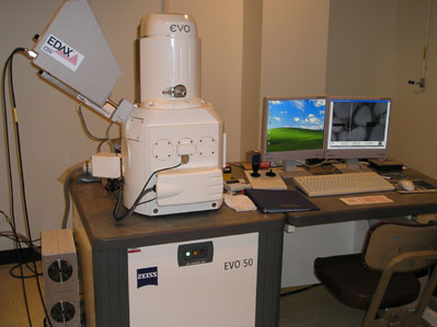 Zeiss EVO-50XVP Environmental Scanning Electron Microscope (ESEM) Zeiss EVO-50XVP Environmental Scanning Electron Microscope (ESEM)This is an extended pressure range SEM with a graphical user interface and digital image storage and processing. It has both a secondary electron (SE) detector and a four-quadrant backscattered electron detector (4Q-BSD). Electron acceleration voltages of 0.2 kV to 30 kV are selectable. The ultimate resolution is 3 nm. This instrument has a large multi-ported specimen chamber with a 5-axis motorized sample stage. The vacuum system uses an oil-free turbomolecular pump. In the extended variable pressure (XVP) mode, pressure from 1 - 750 Pa can be selected. The specimen atmosphere can be either air or water. Integrated with the Zeiss ESEM is an EDAX Genesis 2000 energy dispersive spectroscopy (EDS) system. This x-ray microanalysis system can detect all elements with atomic number greater than or equal to 5 and has software for calculating sample composition as well as for elemental mapping and compositional line scans. Click here for more detailed information about the capabilities of the ESEM. Visit the Gallery to see images obtained using the ESEM.
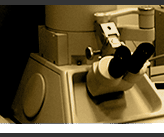
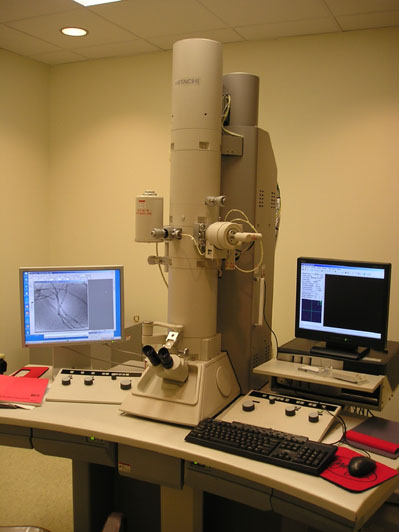 Hitachi H-7600 Transmission Electron Microscope (TEM) Hitachi H-7600 Transmission Electron Microscope (TEM)This TEM has an auto-focus capability with computer-controlled microscope operation (Windows R2000 OS3). Accelerating voltages of 40 kV to 120 kV are selectable. The ultimate resolution is 0.2 nm (lattice image). Samples are mounted on a 3 mm diameter specimen grid. This instrument has an AMT digital CCD system with a bottom-mount camera for acquisition of high-quality digital sample images. Click here for more detailed information about the capabilities of the TEM. Visit the Gallery to see images obtained using the TEM.
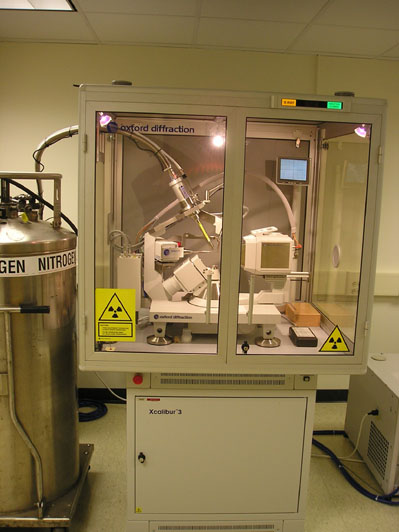 Oxford Diffraction Xcalibur 3 x-ray diffractometer (XRD) Oxford Diffraction Xcalibur 3 x-ray diffractometer (XRD)This is a fully-enclosed single crystal XRD system. It has a Mo/Cu x-ray source with a Sapphire3 CCD detector. The instrument is computer-controlled with CrysAlisTM software for data analysis. Samples can be cooled to temperatures between 90 and 300 K with the Cryojet nitrogen jet cold unit. Renishaw inVia Reflex Micro-Raman
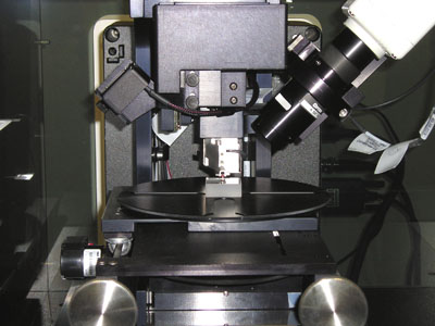
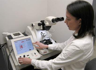
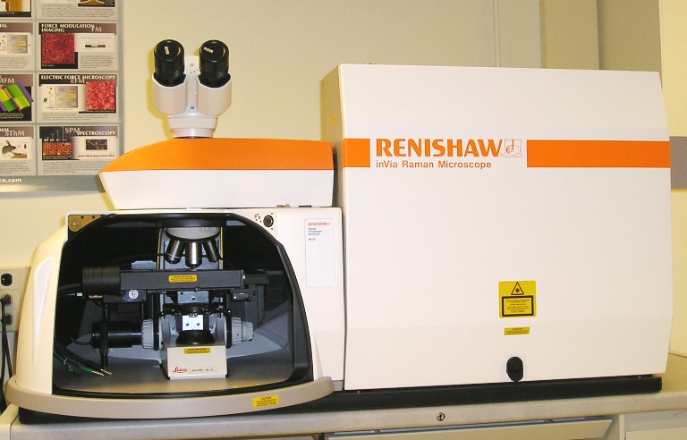 The Renishaw inVia Reflex Micro-Raman is equipped with two laser excitation sources: a near infrared diode laser source (300mW) with an excitation wavelength of 785nm and an Argon ion laser source (25mW) with an excitation wavelength of 514nm. The laser spot size can be varied from 1 µm to 300 µm. Spectra can be obtained using a variety of acquisition modes such as single point scan, line mapping and area mapping. The confocal capabilities of the instrument allow depth profiling with 2.5 µm resolution. The white light image montaging feature allows for large-area images of the sample surface to be captured, which can then be overlaid and compared with Raman spectral data maps of the same region. The instrument is designed for one-click switching between laser sources, with computer-controlled reconfiguration and optimization. Calibration is also automatic, using an internal reference sample. Click here for more detailed information about the capabilities of the Micro-Raman. Veeco Dektak 6M Surface Profiler Leica EM UC6 Ultramicrotome with FC6 Attachment Last updated on October 3, 2013. |